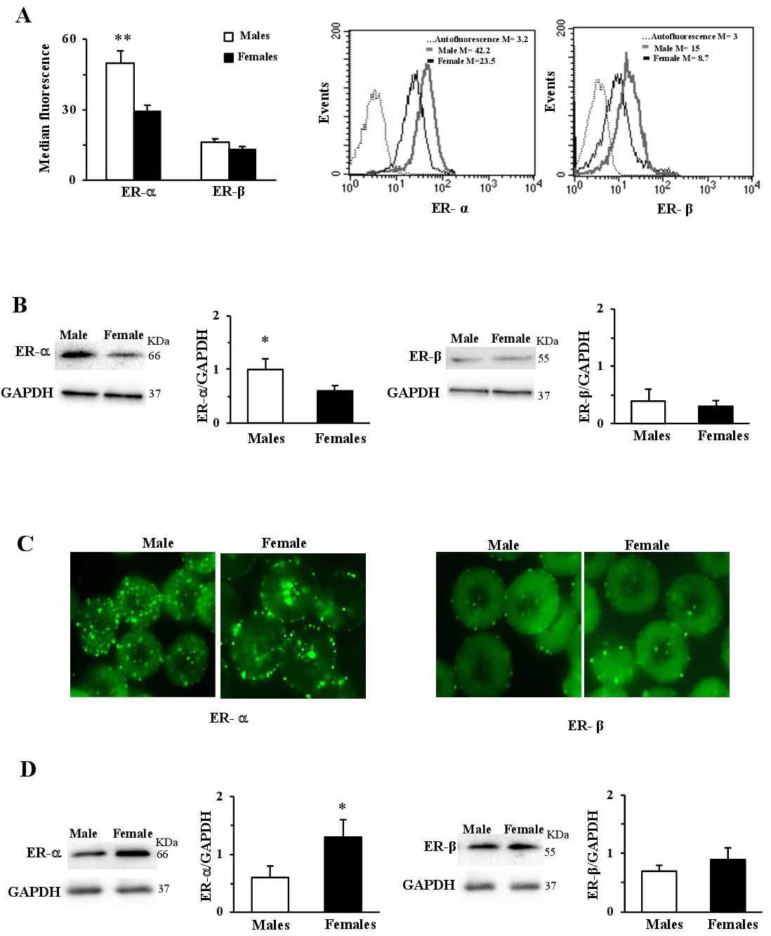
Fig. 1. Estrogen receptors (ERs) exist in human red blood cells and are differently present in males and females. (A, left panel) Cytometric analysis of ER-α and ER-β content in RBCs from males and females. ER-α content was significantly (p<0.001) higher in RBCs from males and ER-β was poorly content in RBCs without any significant difference between the two sexes. Numbers are the median values of fluorescence intensity. (A, right panels) Evaluation of ERs in RBCs from a representative male and a representative female. (B) Representative blotting of ER-α (left panels) and ER-β (right panels), normalized for the GAPDH content, and histograms showing densitometric analysis of three different experiments. The western blotting analysis confirmed the cytometric data (p<0.05). (C, left panels) Micrographs obtained by static cytometry showing ER-α distribution in RBCs from representative male and representative female. (C, right panels) Micrographs obtained by static cytometry showing ER-β distribution in RBCs from a representative male and a representative female. In RBCs from the males, ER-α was mainly localized in the cytoplasm, while in RBCs from the females it was mainly localized in the cell membrane. Conversely, no sex difference was detectable in ER-β distribution. (D) Representative blotting of ER-α and ER-β membrane levels, and densitometry analysis of three different experiments. Membrane ER-α content was higher in RBCs from females with respect to males (p<0.05) (D, left panels), and membrane ER-β content was comparable in RBCs from both sexes (D, right panels). * p<0.05; ** p<0.001.